Advanced Ultrasound Technology for Muscle Strain Diagnosis and Treatment

6F, Building B4,Shenchengtou creative factory life science park, Julongshan A Road, Xiuxin block, Kengzi Street, Pingshan District, Shenzhen,China +86-75586704556 [email protected]

Get a Free Quote

Our representative will contact you soon.
Email
Name
Company Name
Message
0/1000

ultrasound for muscle strain

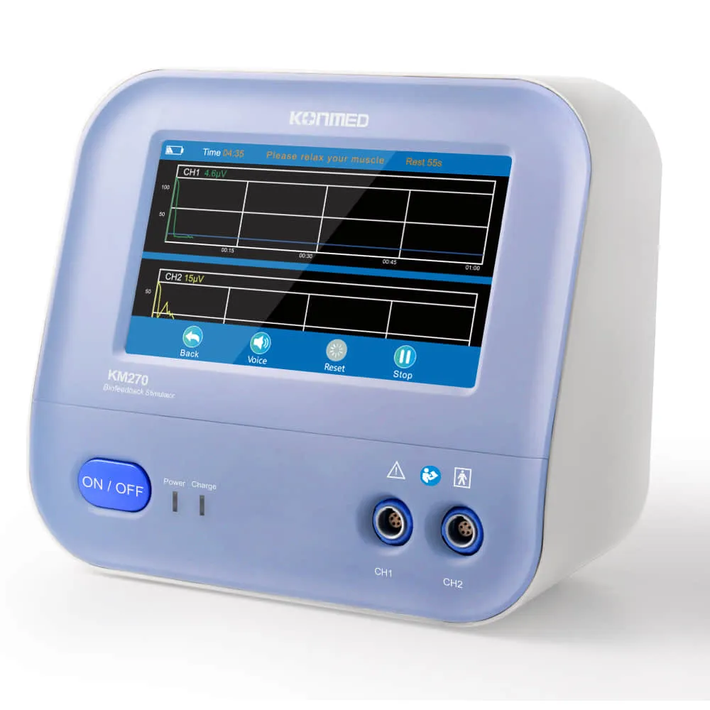
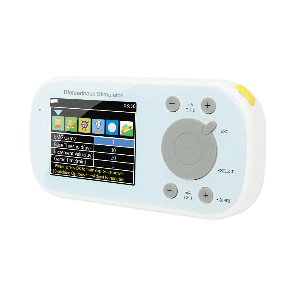
Ultrasound for muscle strain represents a cutting-edge diagnostic and therapeutic tool that has revolutionized the assessment and treatment of musculoskeletal injuries. This non-invasive technology utilizes high-frequency sound waves to create detailed images of soft tissues, allowing healthcare providers to visualize muscle tears, inflammation, and other related injuries in real-time. The technology works by emitting sound waves that bounce off different tissue structures, creating detailed images that can be interpreted immediately on a digital screen. Modern ultrasound devices for muscle strain assessment feature advanced imaging capabilities, including Doppler technology that can assess blood flow patterns within the affected area, helping to identify areas of inflammation and healing. The equipment typically includes a handheld transducer that can be precisely positioned over the injured area, providing multiple viewing angles and depth options for comprehensive assessment. This versatility makes it particularly valuable for examining different muscle groups, from superficial injuries to deeper tissue damage. Healthcare providers can use these images to determine the exact location and severity of muscle strains, enabling them to develop more targeted and effective treatment plans. The immediate availability of results allows for quick decision-making regarding treatment options and return-to-activity timelines.

Popular Products

Ultrasound technology for muscle strain offers numerous compelling advantages that make it an invaluable tool in modern healthcare. First and foremost, its non-invasive nature means patients can undergo examination without any discomfort or recovery time. The real-time imaging capability allows healthcare providers to conduct dynamic assessments, observing how muscles and surrounding tissues move during active motion, which is crucial for accurate diagnosis. This immediate feedback helps in determining the extent of injury and monitoring healing progress over time. The technology is extremely safe, using no ionizing radiation, making it suitable for repeated examinations without health risks. Cost-effectiveness is another significant advantage, as ultrasound examinations are generally more affordable than other imaging modalities like MRI or CT scans. The portability of modern ultrasound devices enables point-of-care assessment, meaning athletes and patients can receive immediate evaluation in various settings, from sports facilities to physical therapy clinics. The high-resolution imaging capabilities allow for detailed visualization of even minor muscle tears and strains, helping healthcare providers make more accurate diagnoses and develop precise treatment plans. The technology also facilitates better patient education, as practitioners can show patients their injury in real-time, improving understanding and compliance with treatment protocols. Additionally, ultrasound guidance can be used for therapeutic interventions, such as precise needle placement for injections or identifying specific areas requiring targeted treatment.

Latest News

How to perform neuromuscular and motor function rehabilitation and relieve pain? Try biofeedback therapy now?

21

Feb

How to perform neuromuscular and motor function rehabilitation and relieve pain? Try biofeedback therapy now?

View More
Biofeedback electrical stimulation helps improve female pelvic floor dysfunction?

21

Feb

Biofeedback electrical stimulation helps improve female pelvic floor dysfunction?

View More
Let you learn about the role of electromyographic biofeedback in improving motor control and quality of life in stroke patients.

21

Feb

Let you learn about the role of electromyographic biofeedback in improving motor control and quality of life in stroke patients.

View More
Vaginal relaxation, frequent urination, urgency, urinary incontinence, sexual dysfunction? Pelvic floor electrical stimu

21

Feb

Vaginal relaxation, frequent urination, urgency, urinary incontinence, sexual dysfunction? Pelvic floor electrical stimu

View More

Get a Free Quote

Our representative will contact you soon.
Email
Name
Company Name
Message
0/1000

ultrasound for muscle strain

Advanced Real-Time Imaging Technology

Advanced Real-Time Imaging Technology

The ultrasound system for muscle strain incorporates state-of-the-art imaging technology that provides unprecedented clarity and detail in real-time visualization. This advanced feature utilizes multi-frequency transducers that can be adjusted to optimize image quality for different tissue depths and types. The system processes thousands of data points per second to create highly detailed images that can distinguish between different types of soft tissue, including muscle fibers, tendons, and surrounding structures. The real-time nature of the imaging allows healthcare providers to observe tissue movement dynamically, which is crucial for understanding how the injury affects muscle function during various movements. This capability is enhanced by color Doppler technology that can detect and display blood flow patterns, helping to identify areas of inflammation and assess healing progress. The system also includes advanced image processing algorithms that reduce noise and enhance contrast, resulting in clearer, more detailed images that aid in accurate diagnosis.
Comprehensive Diagnostic Capabilities

Comprehensive Diagnostic Capabilities

The diagnostic capabilities of ultrasound for muscle strain extend far beyond simple visualization. The system enables healthcare providers to perform detailed measurements of tissue damage, assess the extent of inflammation, and evaluate the integrity of surrounding structures. Multiple imaging modes allow for both broad overview scans and detailed close-up examinations of specific areas of concern. The technology can detect subtle changes in tissue structure that might indicate early signs of injury or healing, helping to prevent further damage and optimize recovery timing. The system includes specialized presets for different muscle groups and injury types, ensuring consistent and reliable imaging across various clinical scenarios. Advanced measurement tools allow for precise quantification of tear size, muscle thickness, and other relevant parameters, providing objective data for tracking recovery progress. This comprehensive diagnostic approach helps healthcare providers make more informed decisions about treatment strategies and return-to-activity timing.
User-Friendly Interface and Workflow Integration

User-Friendly Interface and Workflow Integration

The ultrasound system features an intuitive user interface designed to streamline the examination process and enhance clinical workflow efficiency. The system incorporates touch-screen controls and customizable presets that allow healthcare providers to quickly adjust settings for different examination types. Image optimization tools automatically adjust parameters based on the selected examination type, reducing the time needed for manual adjustments. The system includes integrated patient management features that allow for easy storage and retrieval of previous examinations, facilitating comparison of images over time to track healing progress. Data export capabilities enable seamless integration with electronic health records and other clinical information systems. The interface also includes educational resources and reference materials that can be accessed during examinations to assist with image interpretation and diagnostic decision-making. These user-friendly features combine to create a more efficient and effective diagnostic process, ultimately benefiting both healthcare providers and patients.